Rapid lateral-flow tests for sickle cell disease have been commercially available for several years. The chemistry is mature: the most-cited products correctly distinguish HbAA, HbAS, and HbSS from a small whole-blood sample with sensitivity and specificity that, in published comparison studies, sit comfortably above 95% against gold-standard hemoglobin electrophoresis. The chemistry is not the rate-limiting step in newborn screening programs in low-resource settings. The interpretation step often is.
That is where on-device computer vision has begun to make a measurable difference. Several recent studies, plus a growing body of program-level data from screening sites in West and Central Africa, have shown that smartphone-based image capture and machine-learning interpretation can reduce the rate of misread results, document the chain of custody, and tie a positive screen to a patient record without the test cassette ever leaving the screening site.
What the human eye actually misses
A trained nurse reading a sickle cell rapid test in good light, with a fresh sample and a clean cassette, gets the right answer the vast majority of the time. The misreads cluster around predictable conditions:
- Faint test lines. Lines that are just barely visible at the read window can be called negative when they are actually weak positives, particularly in heterozygote (HbAS) samples.
- Ambient lighting variation. Outdoor screening sites and clinics with intermittent power produce inconsistent lighting that affects perceived line intensity.
- Reader fatigue. A nurse reading the 200th cassette of a clinic day reads differently than one reading the 5th.
- Cassette orientation and timing. Most rapid test windows are interpretable in a finite time window. Reads outside that window can drift.
An interpretation model running on a phone does not solve the chemistry problems. It does, however, neutralize most of the operational ones. It captures lighting metadata, normalizes for it, applies a uniform decision threshold across all reads, timestamps the capture, and produces a result that does not depend on the alertness of the reader at hour seven of a clinic.
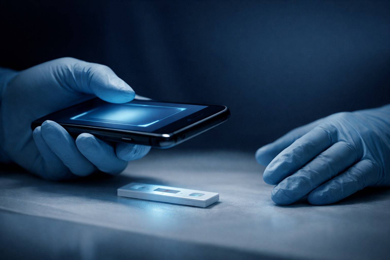
What an AI-assisted workflow actually looks like
The general pattern, across the academic prototypes and the commercial products entering the space, is consistent:
- The cassette runs the way it always has. Sample applied, buffer added, wait the specified time.
- The reader holds a phone over the cassette. An overlay on the phone screen aligns the cassette and confirms the read window is in frame.
- The phone captures one or several images, normalized for white balance and exposure.
- An on-device model classifies the result (HbAA, HbAS, HbSS, invalid) and returns a confidence score.
- The result and the captured image are stored locally and synced to a screening registry when connectivity returns.
The model does not replace the trained nurse. It standardizes the read, time-stamps it, and gives the program a chain of custody that a paper log cannot.
The peer-reviewed evidence
A growing body of work has evaluated smartphone-based interpretation of sickle cell rapid tests, including studies indexed in PubMed by groups working with the World Health Organization on point-of-care diagnostics for sickle cell disease in sub-Saharan Africa. Reported agreement between AI interpretation and expert reader consensus has generally exceeded 95%, with the largest gains observed in heterozygote samples where line intensity is lowest.
Reported limitations are also consistent: cassette-design dependence (a model trained on one manufacturer's cassette does not necessarily transfer to another), lighting-condition robustness in extreme environments, and the operational reality that any model needs an update pathway when a manufacturer changes the cassette housing or print color.
What to ask a vendor
For programs evaluating an AI-assisted rapid sickle cell workflow, four questions tend to surface the real differences:
What cassette did the model train on, and on what sample population. A model trained on adult venous blood may behave differently on heel-stick newborn samples. Ask for the training data demographics.
What is the on-device confidence threshold, and what happens to low-confidence reads. The interesting answer is "they get flagged for human review and cached locally," not "they get reported as a definitive result."
How is the model updated. A model that cannot be re-trained when the cassette housing changes, or when a new printed reference key is added, becomes obsolete the next time the manufacturer rotates packaging.
What is the offline behavior. Many target environments have intermittent connectivity. The model needs to run on-device and the registry needs to sync when bandwidth returns.
Where this points
Our team has spent considerable design time on rapid screening for sickle cell disease, including work on printed reference keys that are intended to make the visual interpretation step more reliable for both human and machine readers. The combination of a well-designed cassette, a printed interpretation reference, and an on-device model is, in our view, the realistic frontier for field diagnostics in low-resource settings. None of the three components alone is sufficient. The three together produce a screen that a clinic without a hematology lab can run, document, and trust.
The chemistry was solved. The next decade of progress in field diagnostics will be measured in how reliably that chemistry can be read, recorded, and acted on by a clinic that does not look anything like a reference lab.